Scientists from the National University of Singapore (NUS) have developed a novel nanodiamond-based contrast agent that improves visualisation of liver cancer tumours. Better and more sensitive imaging helps in detection of liver cancer and is crucial for planning for treatment.
A contrast agent is a chemical "dye" used to enhance the visibility of internal body structures in magnetic resonance imaging (MRI) for cancer diagnosis and to track the progress of patients after treatment.
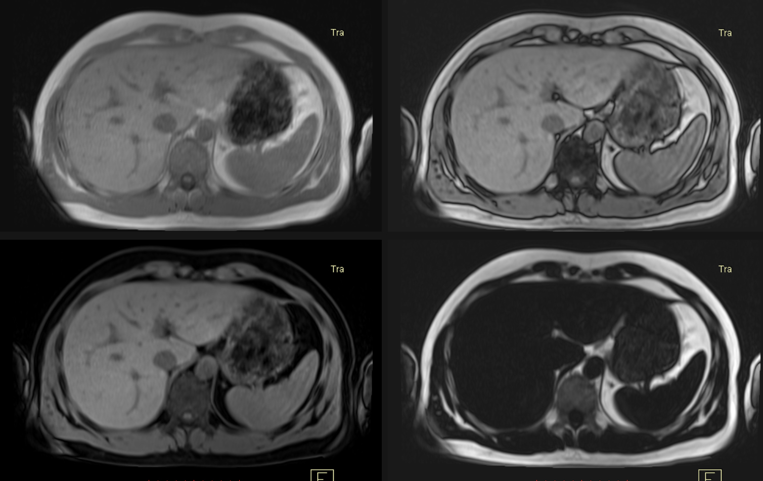
Currently, there are two modes of MRI imaging – T1-weighted and T2-weighted imaging. Patients are often given contrast agents to improve imaging quality.
However, each imaging mode requires a specific class of contrast agent which cannot be used together. This poses a greater challenge in the diagnosis of liver cancer, where T2-weighted imaging is still not considered reliable, and both T1- and T2-weighted imaging can be confounded by tumour vascularity.
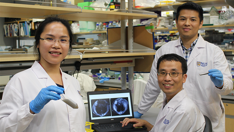
A research team led by Assistant Professor Edward Chow, Principal Investigator from the Cancer Science Institute of Singapore at NUS and Department of Pharmacology at NUS Yong Loo Lin of Medicine, has developed a dual-mode contrast agent which enables clearer and more accurate images of tumours to be obtained in both T1- and T2-weighted MRI scans, and with lower dosages of contrast agent.
“Our experiments suggest that our dual-mode contrast agent holds great promise in improving imaging for liver cancer," said Assistant Professor Chow. "We are hopeful that this advancement in nanomedicine will lead to safer and more accurate diagnosis of liver cancer. Moving forward, we plan to conduct further pre-clinical safety studies for our contrast agents, with the end goal being clinical implementation. We are also looking into using our contrast agents to improve imaging for glioma and ovarian cancer."
The study was conducted in collaboration with the NUS Comparative Medicine Imaging Facility and the Agency for Science, Technology and Research’s Singapore Bioimaging Consortium. The findings of the study were published in the scientific journal Nanomedicine: Nanotechnology, Biology and Medicine in April 2017.